


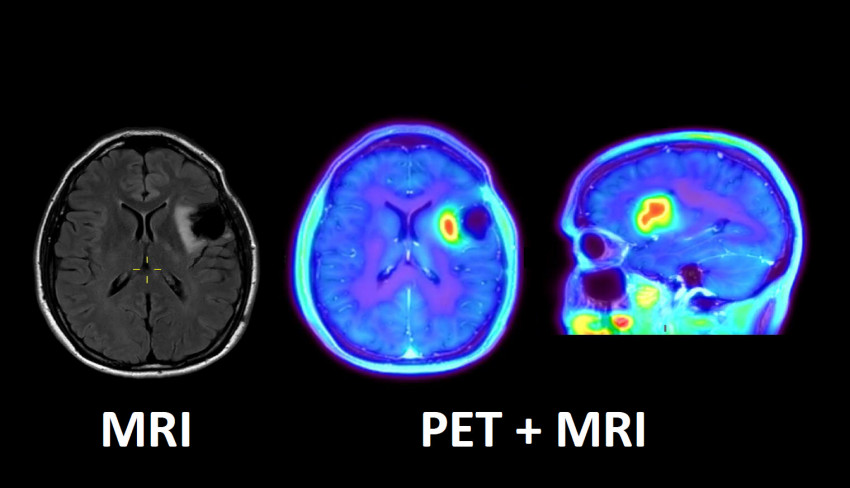
FET (Fluoroethyl-Tyrosine) is an amino acid and is included in regions where many proteins are produced. This is the case, for example, with some malignant brain tumors such as glioblastoma. Using a PET / MRI scanner (both positron emission tomography and magnetic resonance imaging), the uptake of FET in a brain tumor can be accurately measured and imaged. This is important, for example, to determine the best location for a neurosurgical biopsy.
The effect of radiation on brain tumors
Imaging with this new method could also help distinguish between the effects of radiation and the growth of a tumor. The purpose of the radiation is to shrink the tumor, but surrounding tissues can also respond to the radiation. With an MRI, it is not always possible to distinguish between a change in the surrounding tissue and any tumor growth. The new FET-PET / MRI is mainly requested for patients who have doubts about the effect of the treatment.
Previously, patients for this study were referred to the PET Research Center in Jülich, Germany. thats it Now served in Maastricht The OncoZON region (Limburg and the northeastern part of Brabant) has direct benefits for UMC + patients:
- Treatment is closer to home, thus less travel time, more accurate and faster diagnostics and thus faster treatment.
- The patient needs to be examined only once, instead of twice in the past.
Close cooperation
The new FET-PET / MRI research is made possible through close collaboration between the various departments at Maastricht UMC +: Imaging, Neurology, Neurosurgery, Radiation Therapy and Medical Oncology, and with the support of the Board of Directors of the Oncology Center and the Brain / Neurology Center.
The novel method for imaging a brain tumor as closely as possible is being implemented in the Maastricht UMC + hybrid PET / MRI scanner. This scanner combines two imaging technologies in one scanner. With PET (positron emission tomography), certain organ and tissue processes can be visualized with a radioactive material. Magnetic resonance imaging (MRI) produces images via a combination of strong magnetic fields and radio waves.
Maastricht UMC + is currently – next door to Erasmus MC in Rotterdam – the only center in the Netherlands with a built-in PET / MRI scanner. It is also one of the first clinics to use this technology not only for research but also for patient care.
New survey equipment
A few years ago, UMC + Maastricht took over Digital PET / PT scanner In use reduces patient examination time. A PET / CT scan differs from its predecessors because the part of the body that can be examined once, for example an organ, is larger. This results in better quality, so that small abnormalities, such as metastases and small sources of infection, are easier to see. In addition, the scans take less time.
Opening ceremony 2021
Would you also like to attend the inaugural ICT and Health event on June 17, 2021? Entrance tickets are free, but they are really gone! So do not wait and Register quickly.


Maya Angelou is a contributor to The Cheraw Chronicle, covering a broad range of topics including news, politics, business, technology, sports, entertainment, and lifestyle. She focuses on delivering clear reporting, useful information, and timely coverage of current events that matter to readers. Her work aims to provide balanced perspectives and accessible insights, helping audiences stay informed about developments in their communities and beyond through accurate, reader-focused storytelling.


More Stories
Venus-Jupiter Conjunction 2026 Dazzles Skywatchers Around the World
Crescent Moon Meets Venus and Jupiter in Rare Evening Sky Display This Week
Rare ‘Blood Moon’ Total Lunar Eclipse to Be Visible Across the United States and Beyond